It’s not every week that you get to see a real brain, but some of our MICDS Upper Schoolers have had the opportunity to see and hold both a sheep brain and human brain over the past couple of weeks.
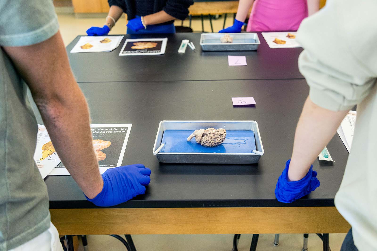
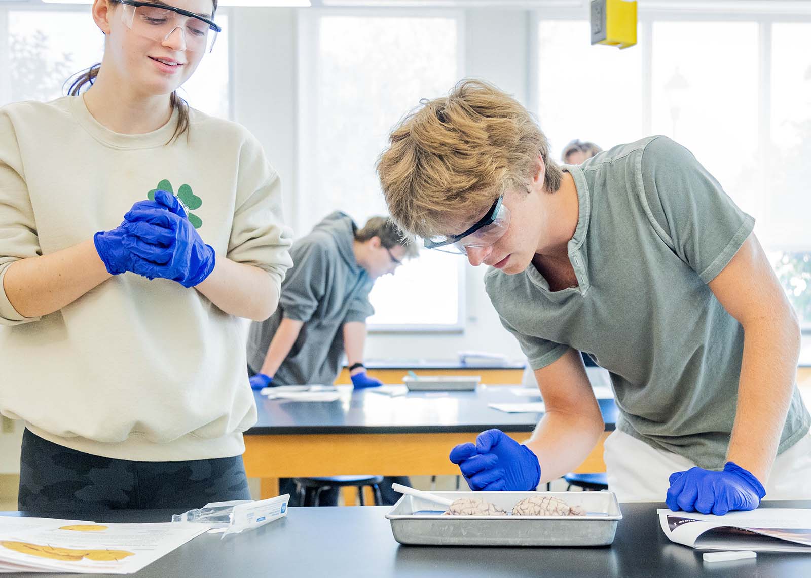
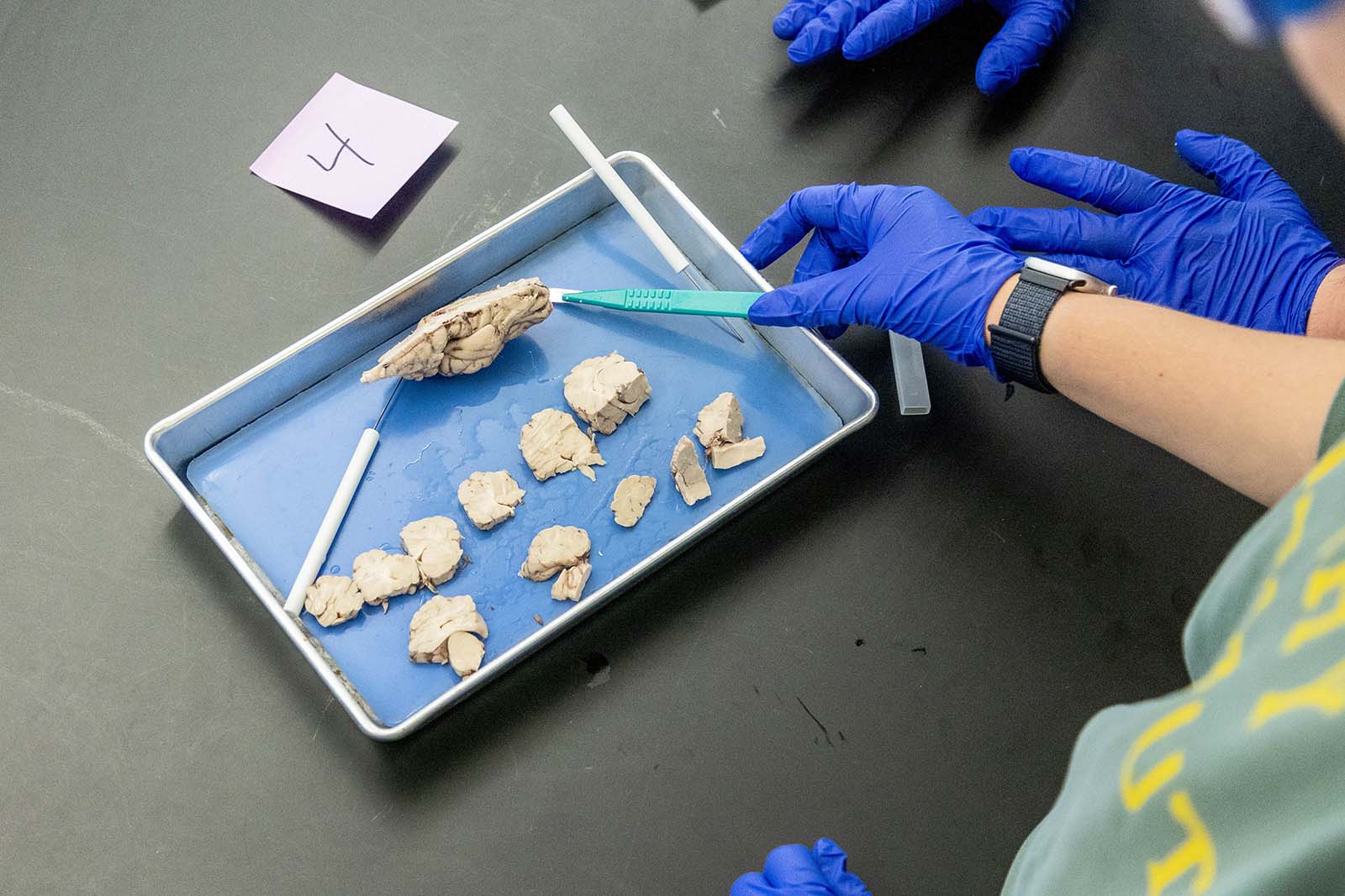
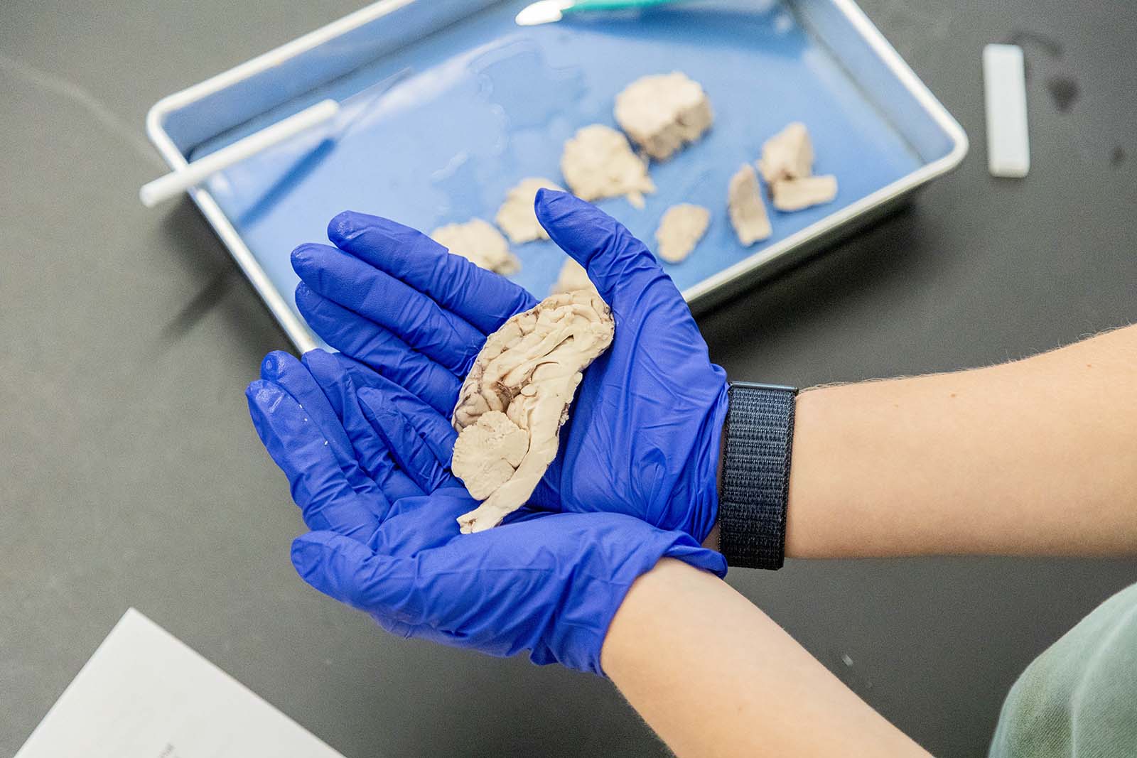
Sheep Brain Dissections
In neuroscience, students spent the semester studying functions of the frontal lobe of the cerebral cortex, such as personality, attitude, mood, emotion, memory, and problem-solving. They learned about the structure and function of neurons that form complex brain circuitry and support brain function. They also studied how signals travel within neurons and across synapses between neurons.
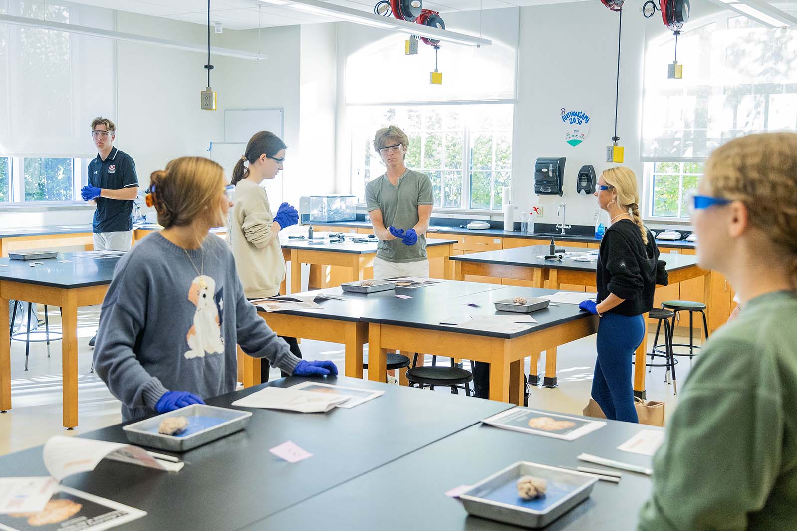
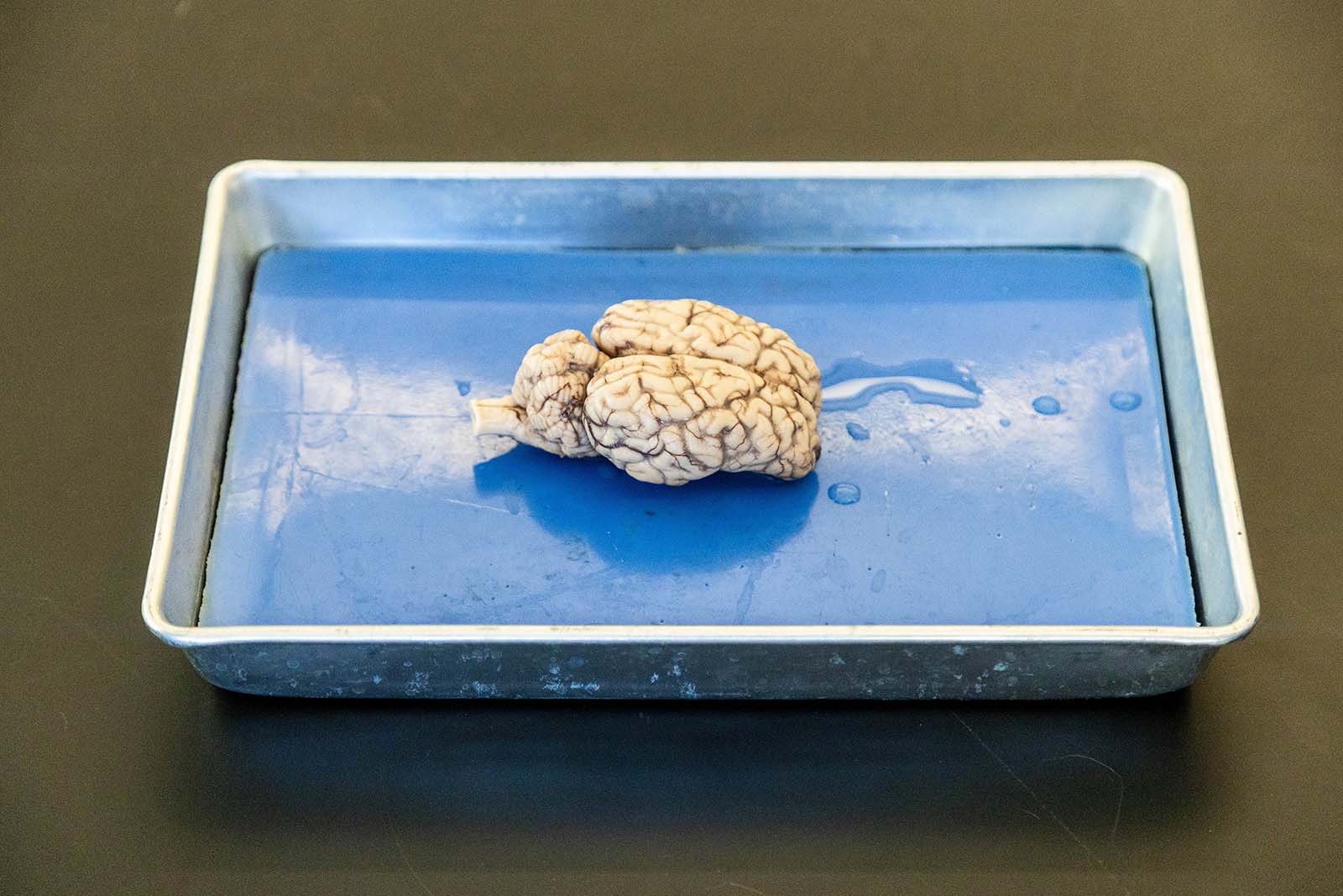
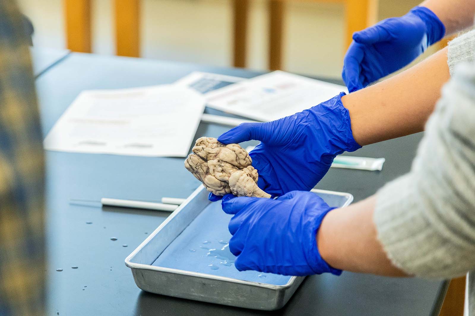
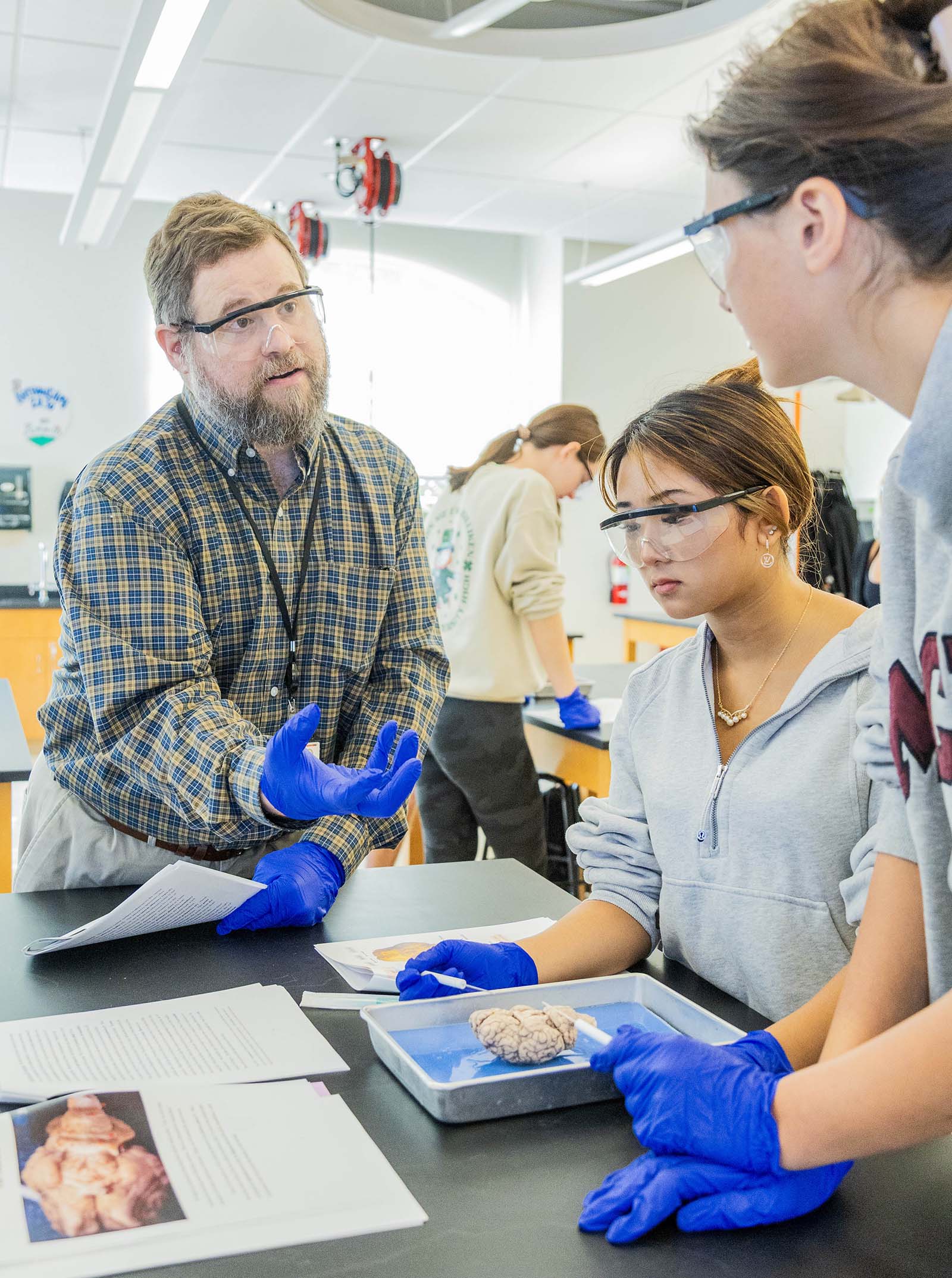
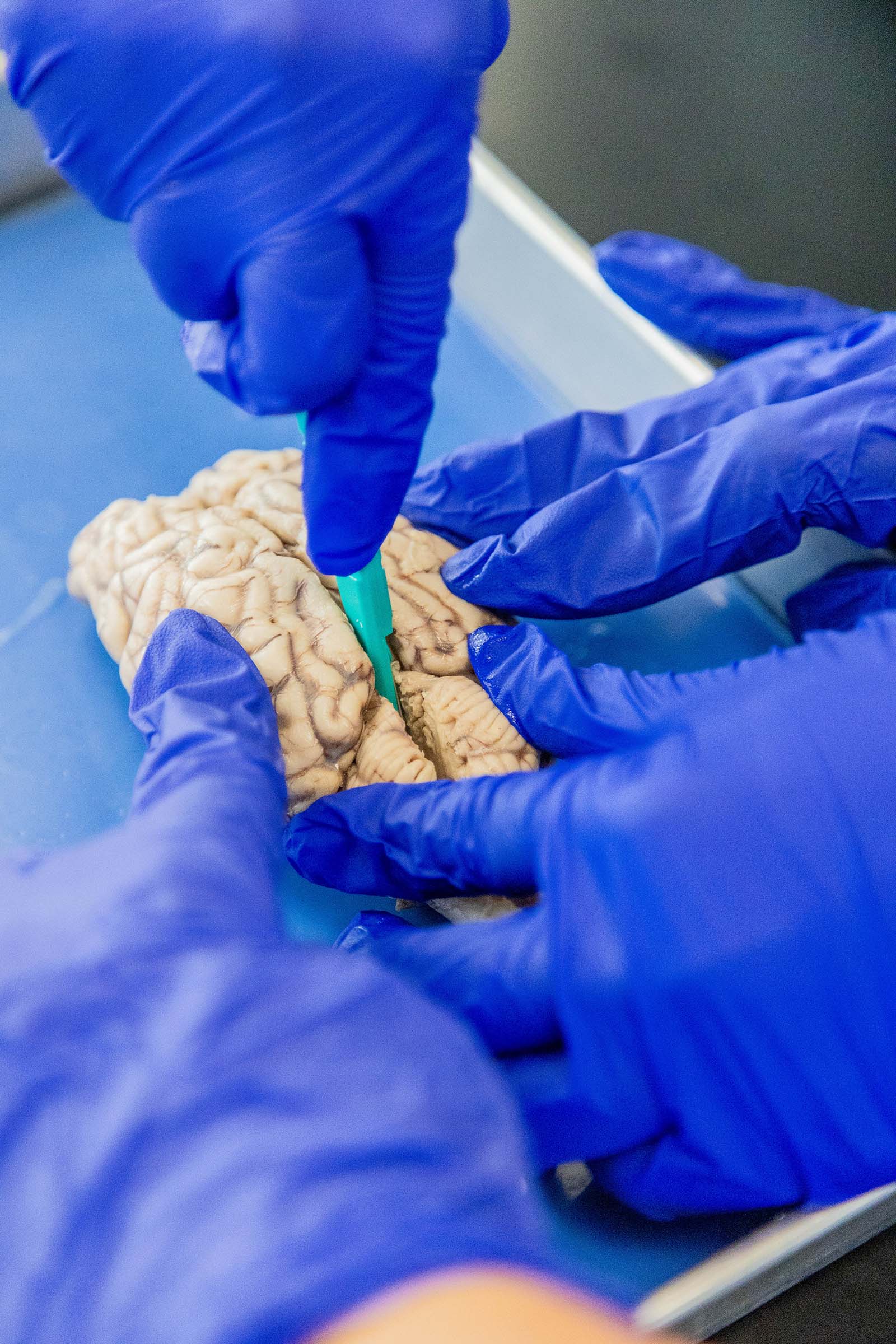
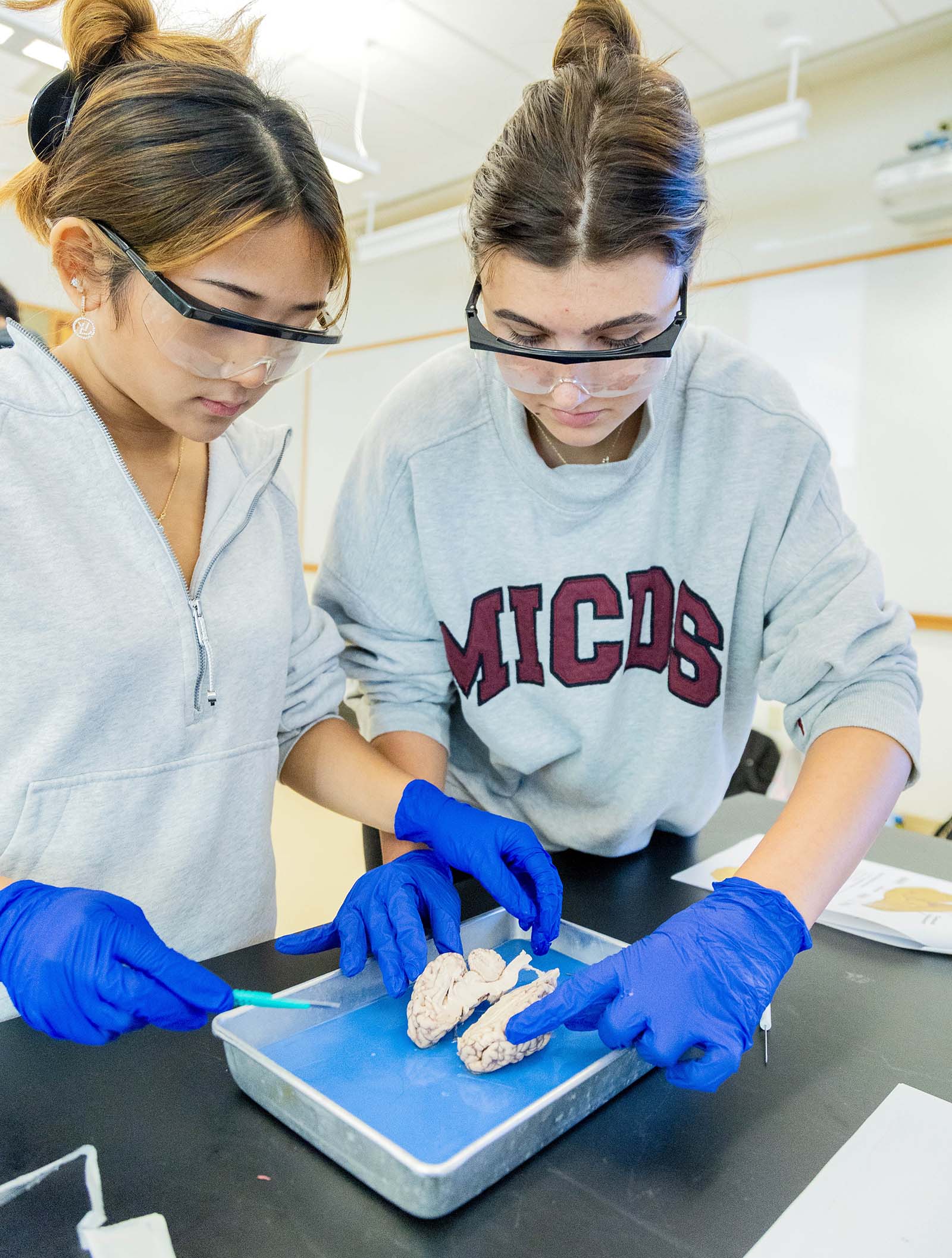
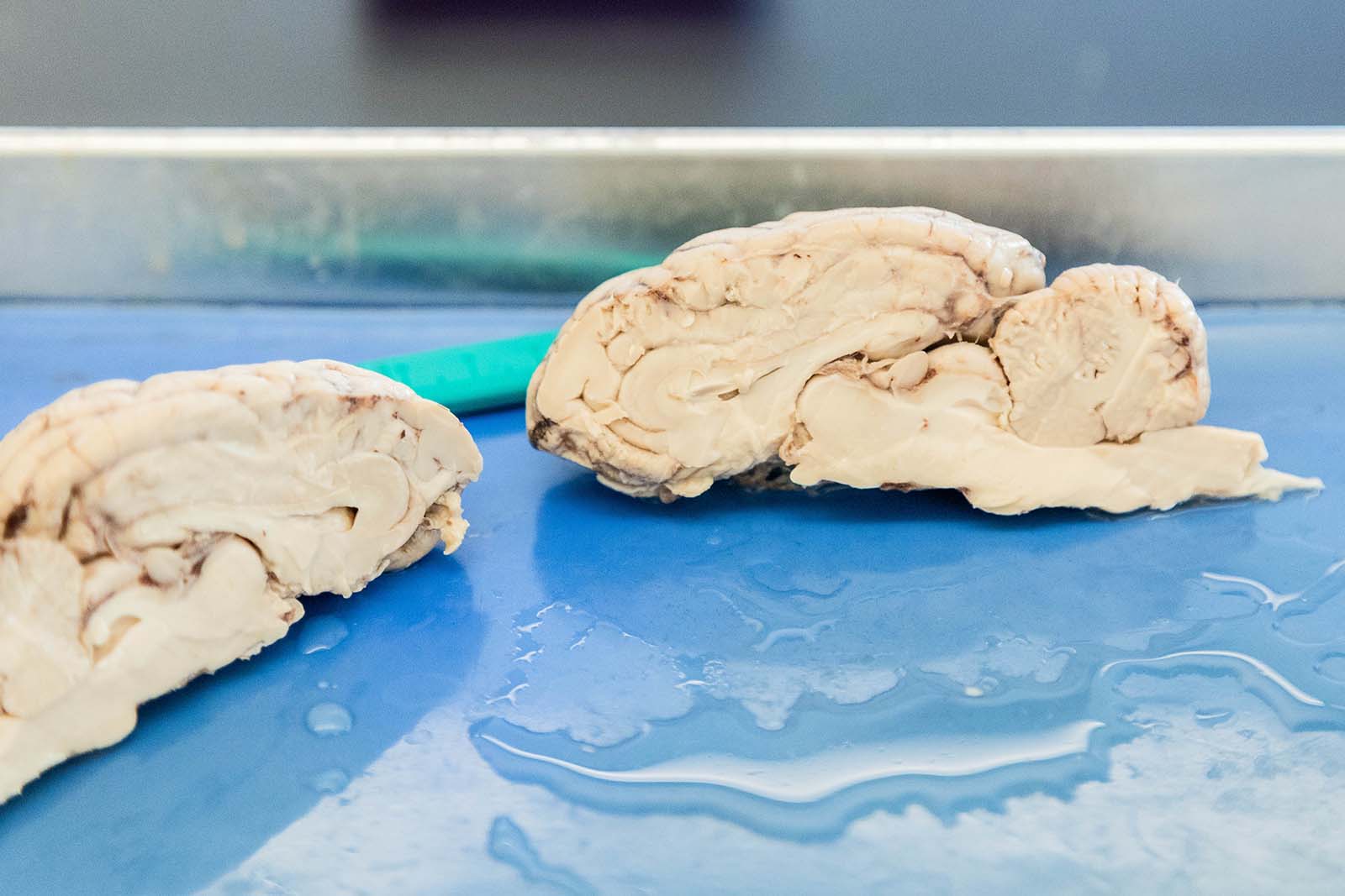
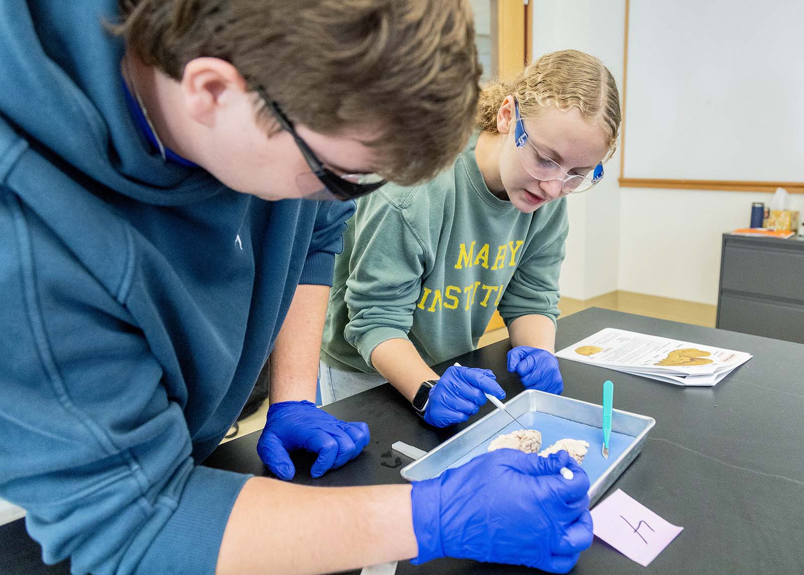
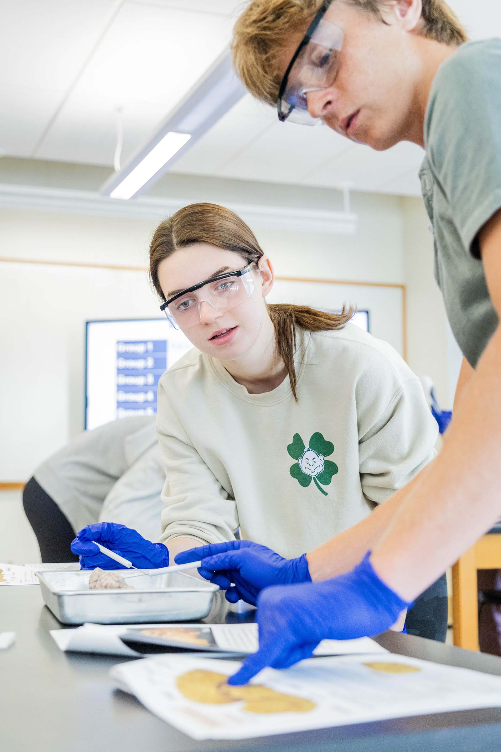
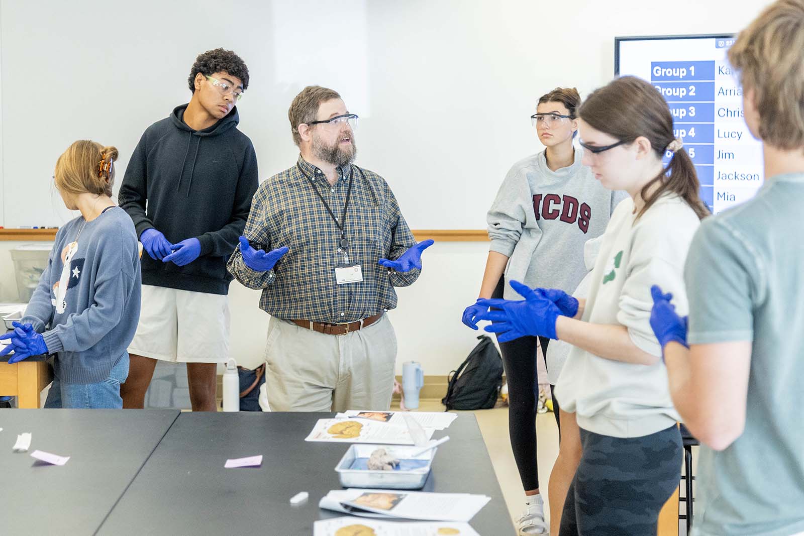
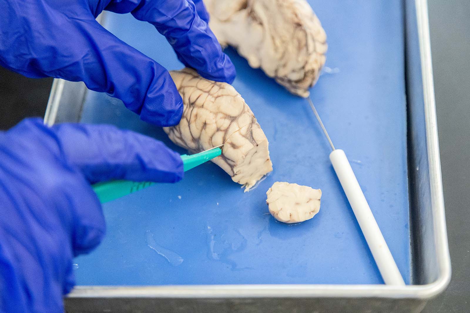
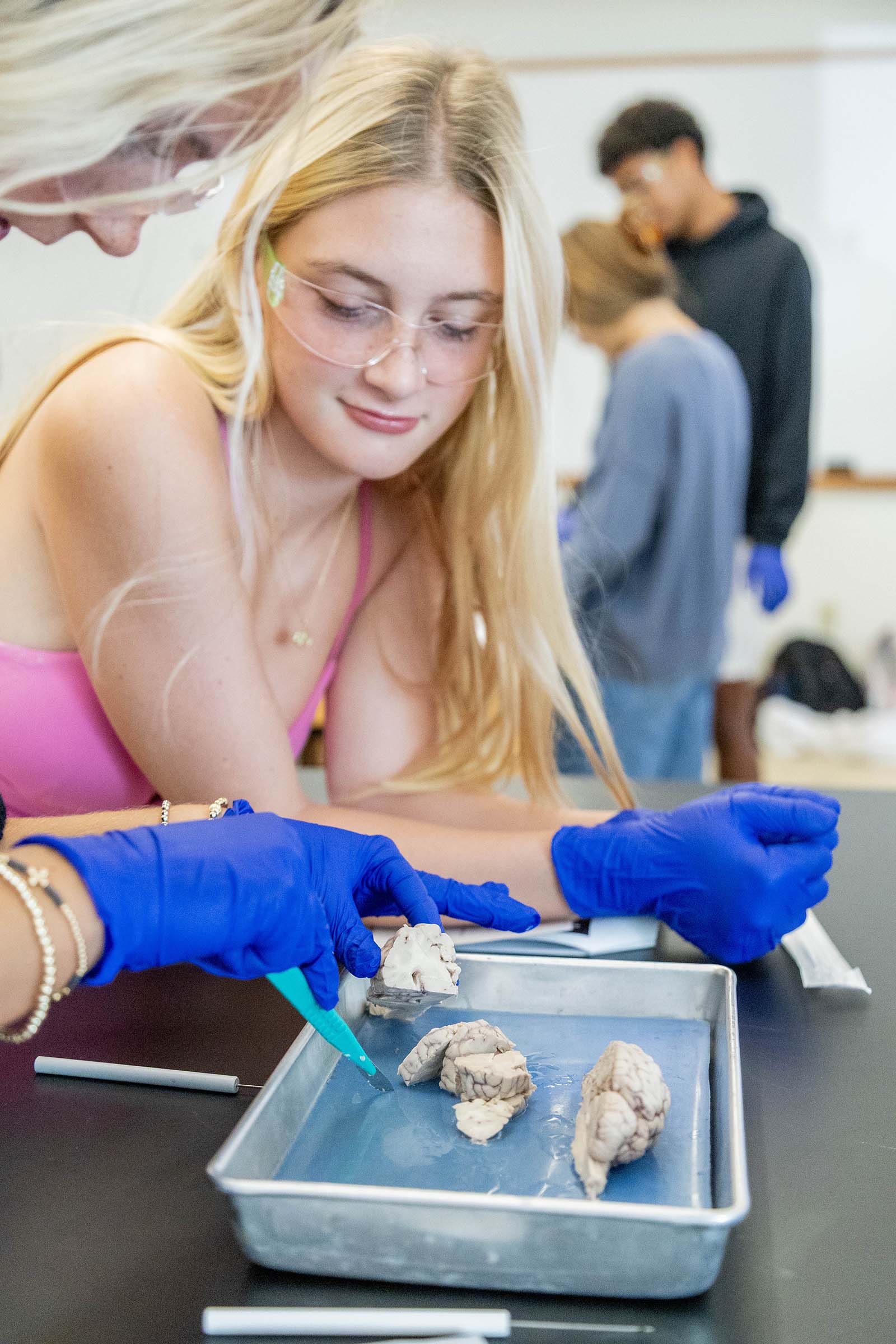
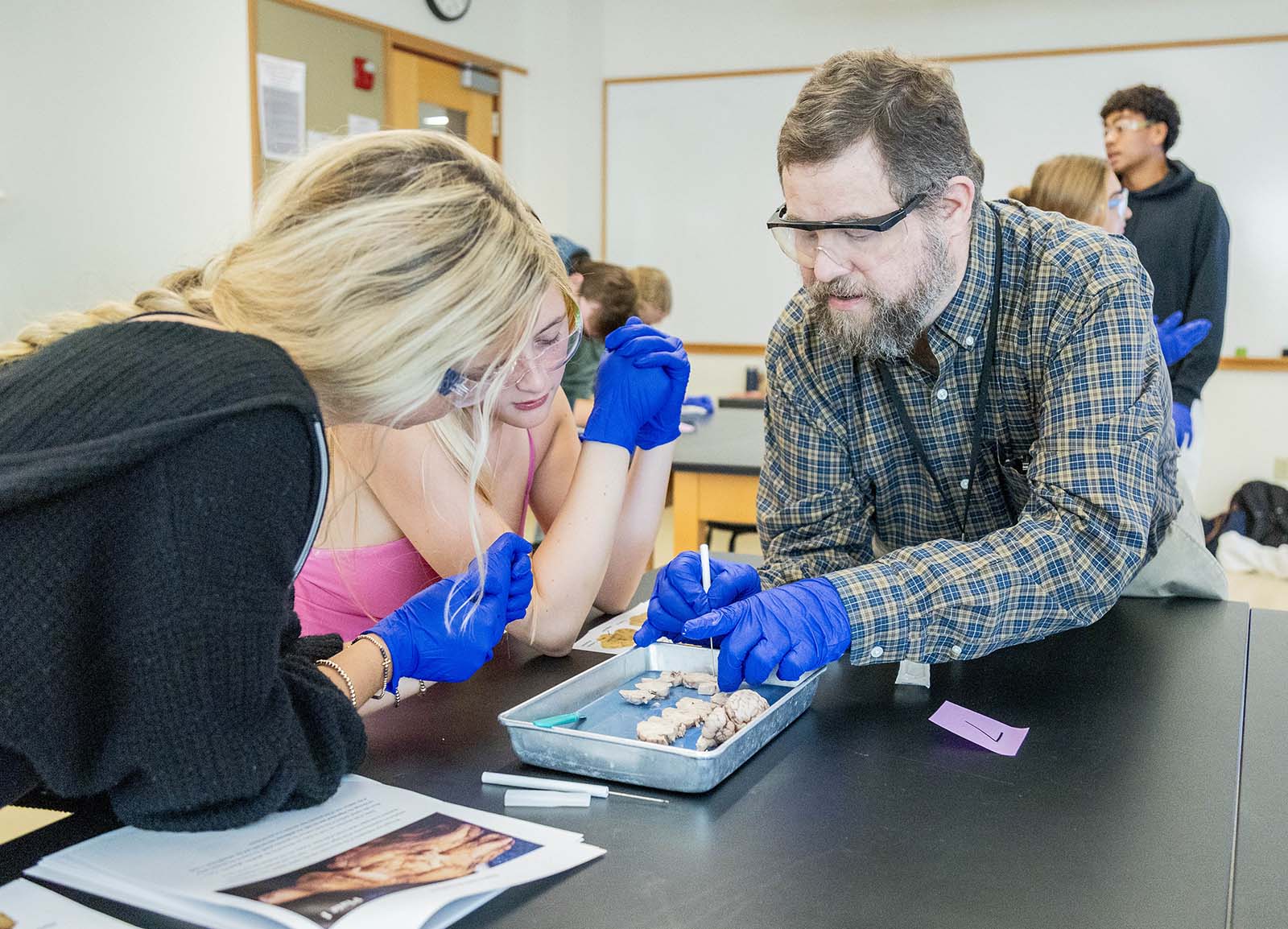
“To prepare for an upcoming presentation with a neurosurgeon, knowing that he would be talking about human brain anatomy and bringing human brain specimens, we dissected sheep brains. Sheep brains have similar structures to human brains, allowing students to learn about brain anatomy in a more experiential setting,” shared Dr. Ethan Graf, Upper School Science Teacher. “This helped students to connect concepts of brain function and neuronal circuit function to where it is all taking place, in the brain itself.”
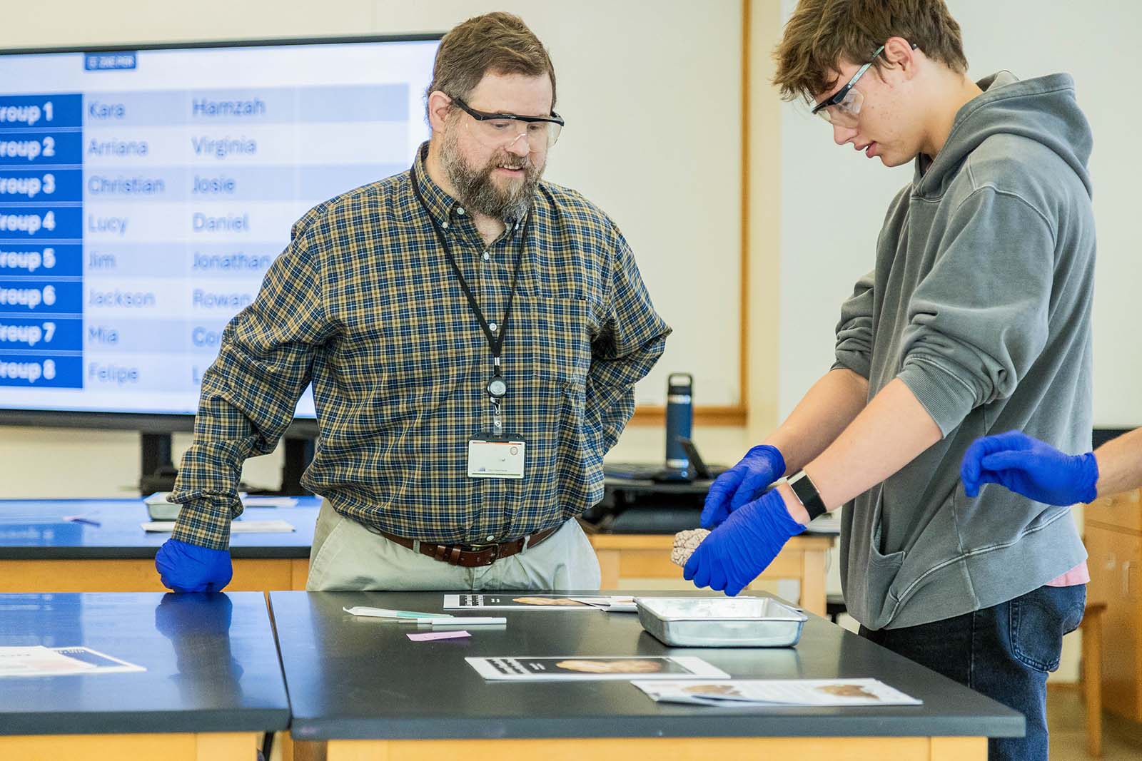
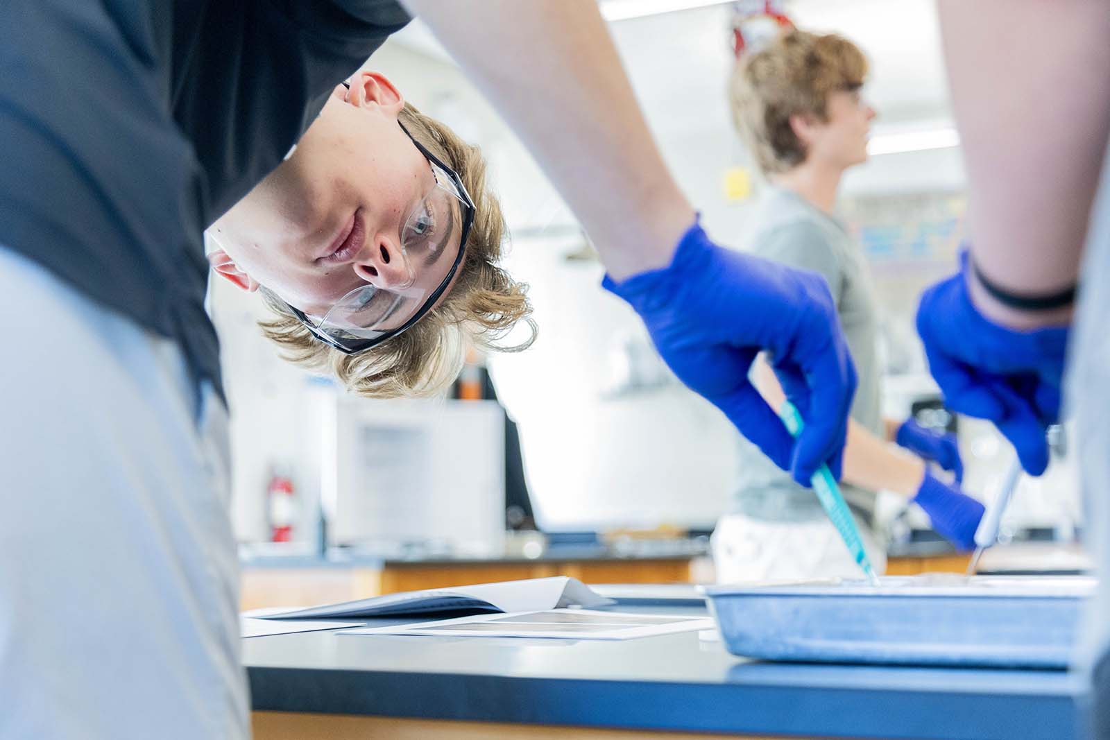
On the day of the dissections, pairs of students were given a sheep brain as Dr. Graf described the different areas of the brain, the functions that occur in each, and the names of the anatomical parts. As students performed the dissections, they were tasked with locating certain parts of the brain and showing Dr. Graf the different structures.
Next week, our AP Psychology students will get to study the sheep brains as well.
Special Guest Visit & Human Brain Lab with Neurosurgeon Dr. Neill Wright
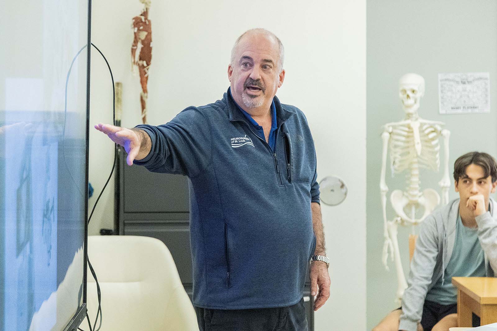
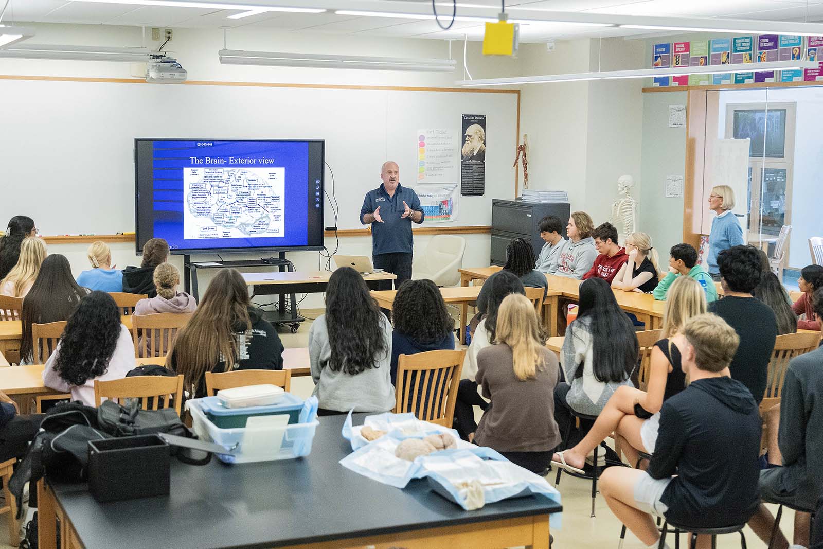
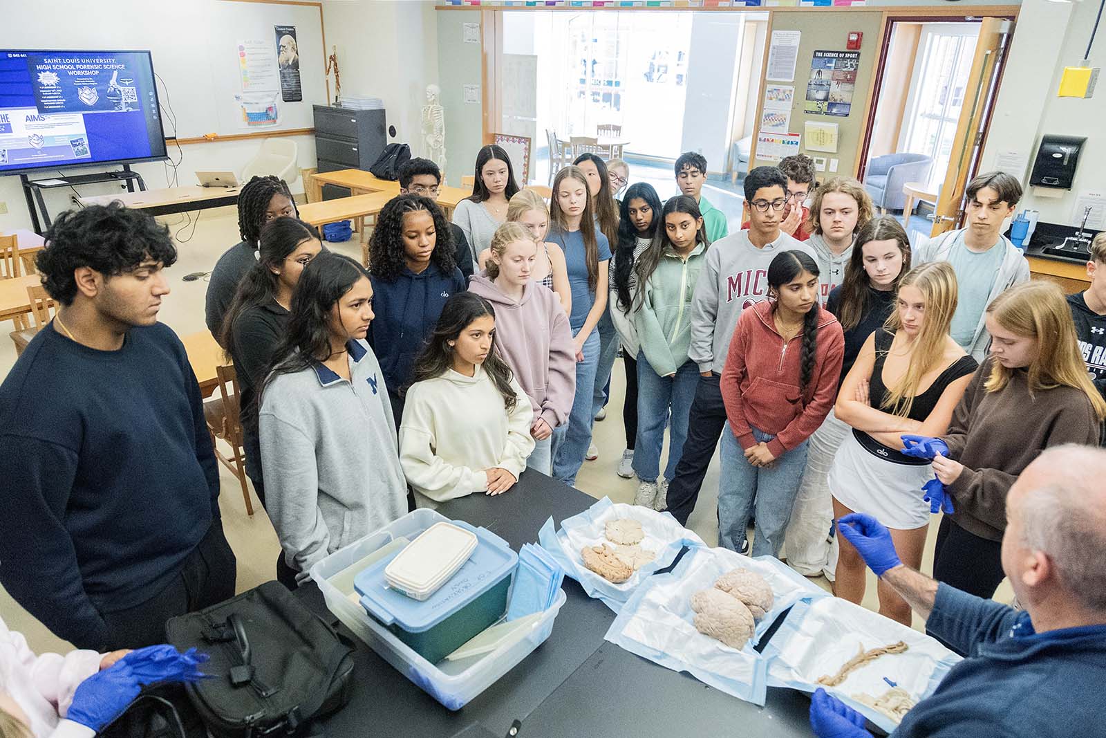
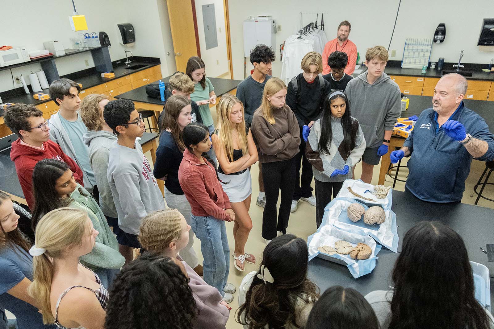
On Monday, October 26, MICDS neuroscience, psychology, and anatomy & physiology students filled STEM 108 to learn from expert neurosurgeon and MICDS parent Dr. Neill M. Wright P ‘25, ’27, ’29. Dr. Wright is the Medical Director at Neurosurgery of St. Louis. He has performed over 2,500 cervical spine surgeries, and his practice also treats craniocervical/craniovertebral junction disorders, atlantoaxial instability, cervical spondylosis, cervical disc herniations, cervical myelopathy, cervical radiculopathy, cervical spondylolisthesis, cervical stenosis, cervical deformity, and tumors of the spinal cord and spine.
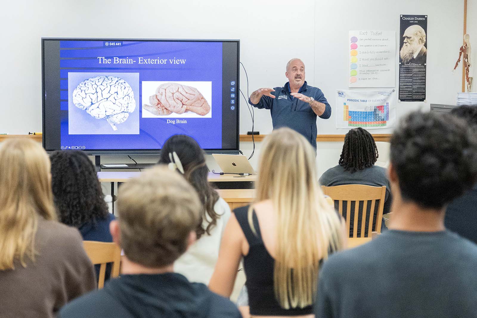
First, Dr. Wright talked about what a neurosurgeon is: a surgeon who specializes in the diagnosis and surgical treatment of disorders of the brain, spinal cord and nerve roots, spine, and peripheral nerves. He walked through his specific journey into neurosurgery, sharing about the lengthy educational path that led him to become a neurosurgeon. Then, knowing that neuroscience students had recently completed their sheep-brain dissections, he compared the human brain to the brains of sheep, dogs, and dolphins. Students learned that the thinking part of the brain happens on the outer surface. The more folds a brain has, the more surface area there is, so there’s more brainpower. The human brain has far more folds than a sheep’s or dog’s brain, but far less surface area than a dolphin’s brain.
In his presentation slides, students saw different views of the brain: axial (horizontal) and sagittal (sideways). Dr. Wright spoke about how important it is to know the function of each area of the brain. Unlike operating on most other parts of the body (like one’s arm), where there is a safe path to the part that needs attention, there’s no safe path to the brain without potentially damaging another part. Neurosurgeons are constantly trying to minimize the side effects of surgery by strategically accessing the surgical site through the area with the least potential for long-term damage. They perform constant risk assessments.
After talking about how the brain normally works, Dr. Wright dove into what neurosurgeons do to help when the brain functions abnormally. He listed a variety of causes that could impact one’s brain: trauma, blood clots, brain bleeds, skull fractures, and more. He also talked about how neurosurgeons help with spinal cord disorders. Dr. Wright explained how some of these issues are treated, what tools they use during surgery, and the counseling involved in discussing the procedure with the patient. He talked about a few client cases and, after warning that the content ahead would be graphic, showed videos to walk through actual surgeries. Students were fascinated to get a glimpse of the actual surgeries and all that goes into a patient’s often life-changing and life-dependent surgery.
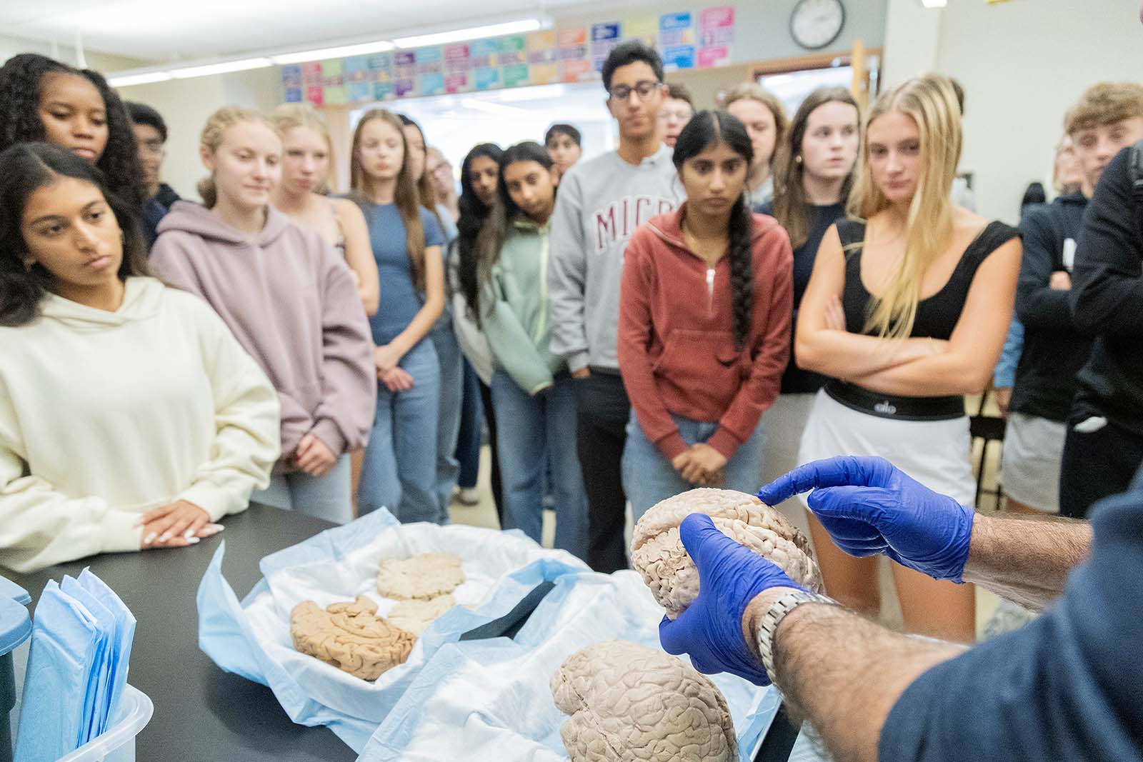
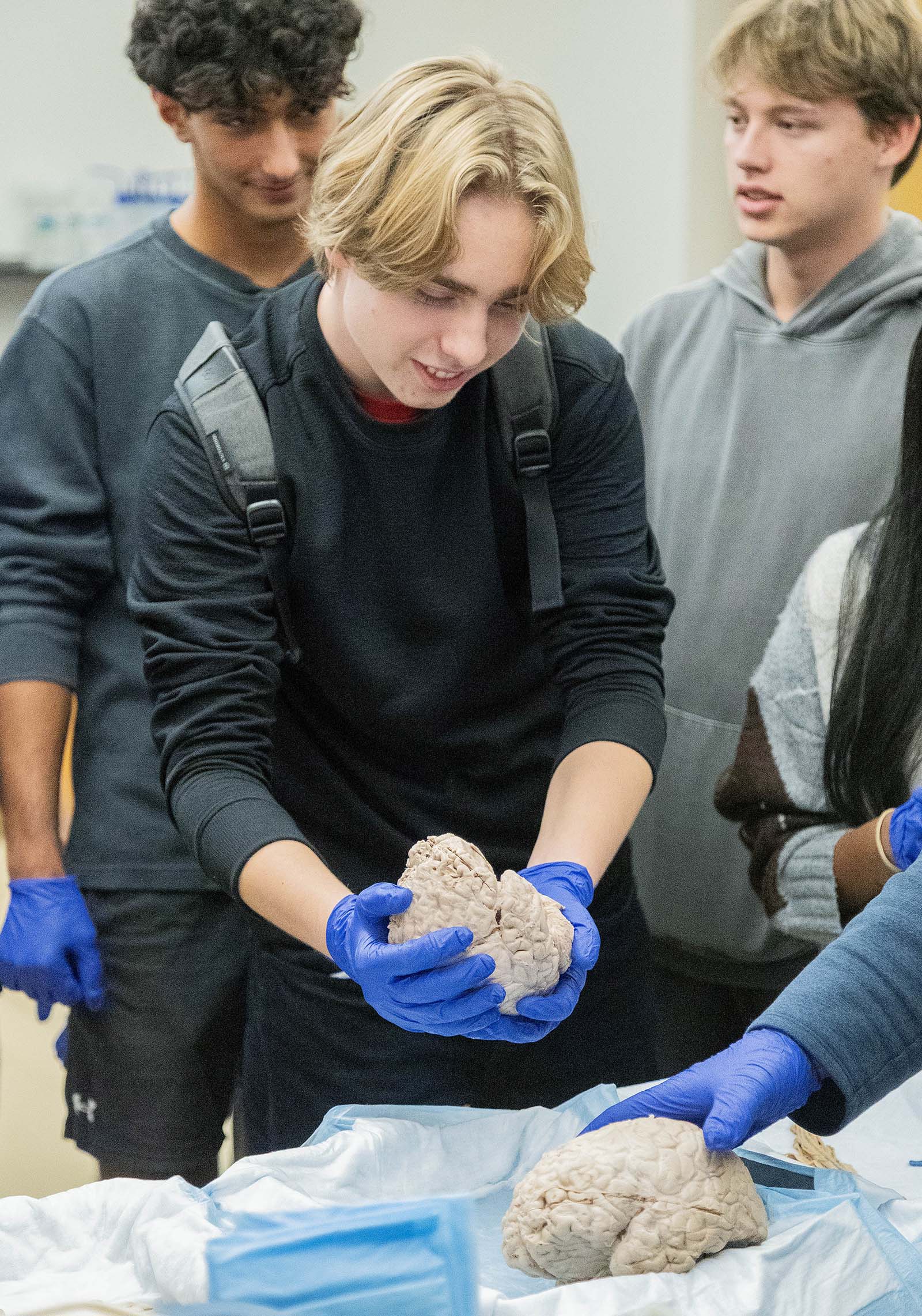
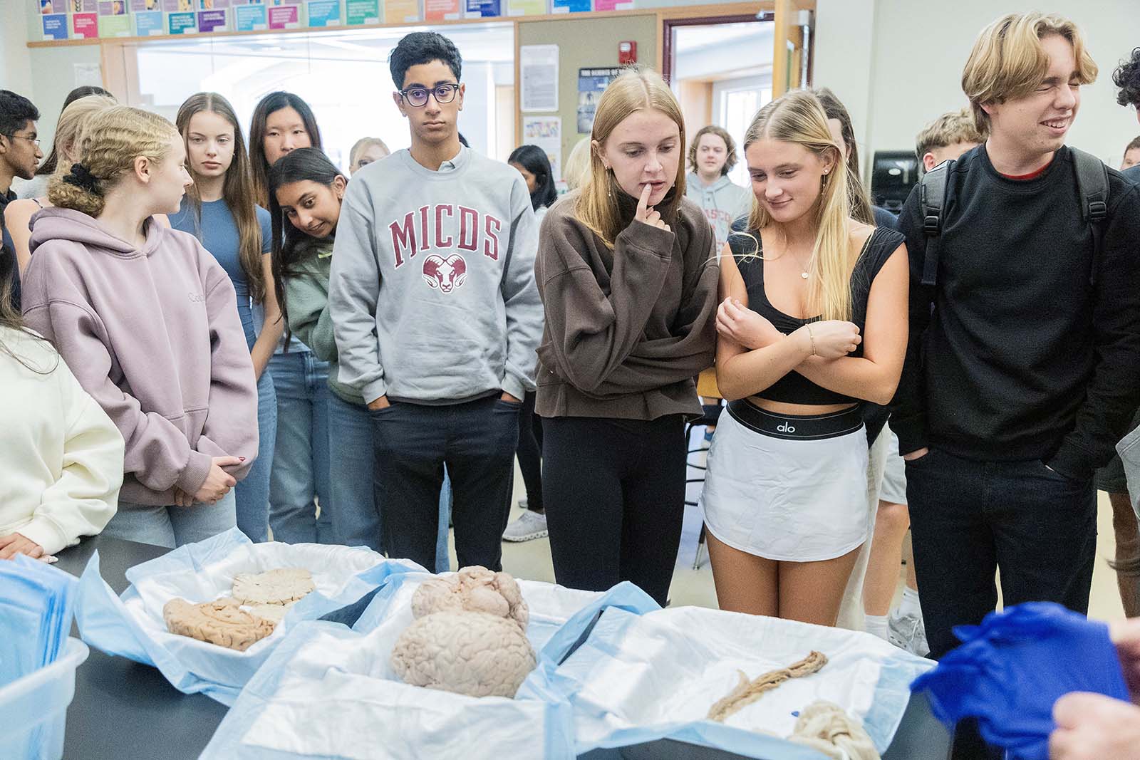
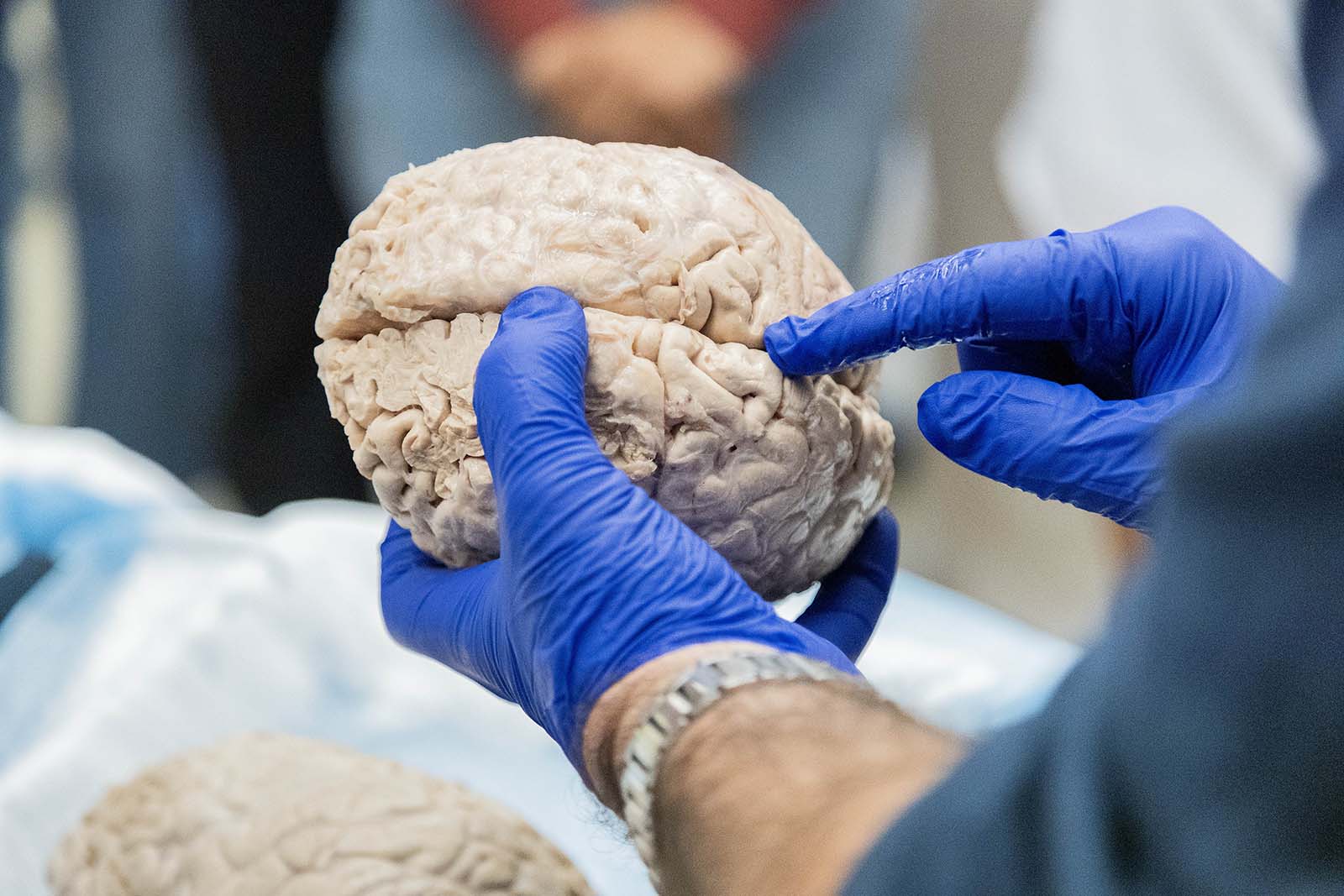
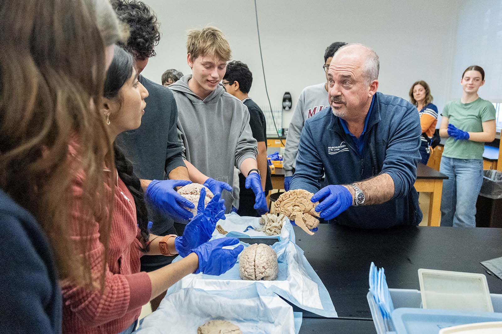
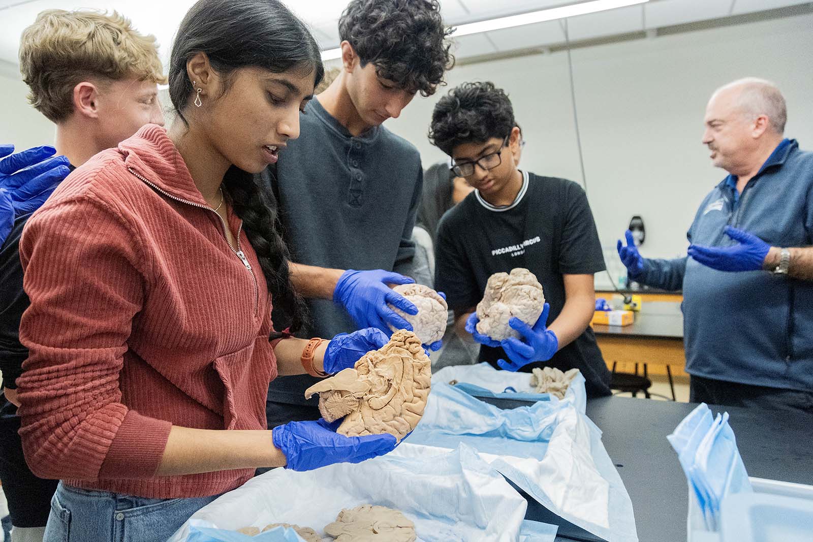
Next, students got to see (and even touch) a real human brain, a dissected spinal cord, skull, and cervical models, which Dr. Wright carefully brought in from the St. Louis University Practical Anatomy Workshop (PASE Lab). The human specimens provided students with a hands-on, interactive learning experience that brought neurology to life. Dr. Wright showed students the various parts of the brain and which parts surgeons avoid. With gloved hands, students could hold the brain to feel how heavy it was or touch the specimens. During the presentation and the brain lab, students asked many questions to better understand Dr. Wright’s work.
In reflecting on the visit with Dr. Wright, Lucy M. ’28 shared, “I thought it was really interesting to see into both the brain and the spinal cord and watch videos on real surgeries that were done. Even though the videos were a little gross, I thought they helped bring together some of the material we have been learning in Neuroscience along with our sheep dissection.”
Arriana T. ’28 added, “I never considered working in surgery in my life, but after watching a few clips and seeing what Dr. Wright could do to support and help others, I thought it was really insightful and interesting. I thought the videos of him performing surgery were fascinating!”
“Dr. Wright’s visit was fantastic,” reflects Paul Zahller, JK-12 Science Department Chair & Upper School Science Teacher. “He provided a unique educational experience for students, including captivating stories and videos from his experience as a neurosurgeon. The specimens provided a once-in-a-lifetime experience for many students to see and touch brain material. We were very fortunate to have Dr. Wright share with us.”
It was a wonderful opportunity to give Upper Schoolers the chance to see what being a neurosurgeon is really like. Thank you very much, Dr. Wright, for sharing your profound experience with our students! Perhaps one day, some of them will become neurosurgeons, psychologists, or physicists who work closely with the brain, just like you!