Science research is alive and well at MICDS, even during the pandemic! Typically, this time of year is when Upper Schoolers present their scientific research on posters during the annual Research Symposium held in Brauer Hall. Of course, the pandemic challenged students to conduct and share their research projects in new and creative ways. Read on to learn about two seniors who have tackled independent studies and research projects this year, advancing their STEM skills before they continue on in college.
“These two research projects demonstrate how the existing curriculum inspires students to seek additional independent research opportunities, make discoveries as agents of science, and develop new skills that they will take to their university experience,” shares JK-12 Science Department Chair Dr. Bob Shaw. “I appreciate the work of the faculty to facilitate these opportunities and experiences that empower our students to pursue their passions in STEM through these specialized research strands at MICDS.”
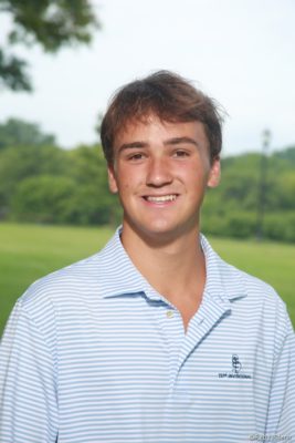
Will Giles ’21 Completes Independent Study Combining Passions for Medicine and Computer Science

For my Independent Study under the guidance of Upper School Math and Computer Science Instructor Ms. Janet Purdy, I updated and modified post-therapeutic documents during my internship at a local orthopedic practice. I further explored this interest by creating a medical diagnostic standard neural network using JAVA. The program essentially gives a preliminary, yet scientifically accurate, diagnosis of particular physical abnormalities using qualitative structured data provided by the user. The program then prints the following information to a file: the probability that the patient has a particular injury, a written description of the patient’s symptoms, potential treatments, and links to medical informational websites.
During the pandemic, I explored machine learning through online Coursera classes in deep learning and found a more pertinent alternative to my initial medical diagnostic program. After taking these classes, I was able to construct convolutional neural network image classification and segmentation models to diagnose brain and chest conditions from x-rays and MRIs. In this class, I identified some of the most common challenges for training convolutional neural networks, such as class imbalance, multitasking, and dataset size, and learned about ways to solve these problems, such as integrating a weighted binary cross-entropy function, in order to improve the accuracy of the model’s predictions. I also learned about common medical evaluation metrics and specifically how to evaluate the accuracy of a model using metrics such as specificity and sensitivity. Towards the end of the course, I used the pre-trained DenseNet architecture to classify conditions in chest x-rays and used the 3-D U-Net model to segment four-dimensional MRI images to identify brain tumors.
The independent study I embarked on during the first semester of this year allowed me to continue studying deep learning models and how machine learning is revolutionizing modern-day medicine. I gained a more thorough understanding of shallow and deep neural networks, how to improve neural networks, how to structure my own machine learning projects, and how artificial intelligence can also be applied to predicting the future health of patients. The above skills were achieved through the course’s combination of core calculus concepts, essential deep learning algorithms, such as gradient descent, logistic regression, and backwards and forward propagation, and through the course’s instruction in the Python coding language. Specifically, my final course project consisted of two parts. During the first part of the project, I constructed a logistic regression neural network that was able to accurately determine if a photo pictured a cat or not. The model analyzed the pixel distributions of the photos and used both forward and backward propagation algorithms to ultimately classify the image as either a 1 or a 0 on a binary scale. A score of a 1 indicates that the photo does picture a cat, and a score of a 0 indicates that the photo does not picture a cat.

During the second project, I constructed a more sophisticated type of machine learning model called a convolutional neural network. This model was able to analyze both two-dimensional and three-dimensional images and similarly classify the contents of the images. The model segmented chest x-rays to determine if the patient had a particular chest deformity, such as a mass or pneumothorax. The model was also able to segment 3-D MRIs to determine if a patient had a brain tumor or not. These models were constructed through both the Deep Learning Specialization and the AI for Medicine Specialization on Coursera.

As I look ahead towards the next four years of my life, I am excited to continue applying what I have already learned in the fields of biology and computer science to more accelerated and complex courses. My work with machine learning has been highly research-dependent, and as I transition into college, I intend to make one of my number one priorities to seek research opportunities. Just as this independent study has required one-on-one collaboration between my faculty sponsor and me, I intend to continue on this path in college and look forward to performing research alongside professors with a focus on medical deep learning models. I’m inspired to learn more and use the tools I will gain in my next four years of higher education to not only continue bettering myself as a STEM student, but also prepare myself for a future in medicine where I’ll be able to help others.
Upon looking back at Will’s work, Ms. Purdy expressed, “His enthusiasm was contagious throughout the entire process. Even during times of challenge, he was thrilled about the possibilities of his project! His perseverance and self-drive pushed him to exceed all expectations while advancing his models far beyond what was originally planned. I simply cannot wait to see what lies ahead for Will!”
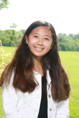
Shudi Wang ’21 Performs Polymerase Chain Reactions, Denaturing Gel Gradient Electrophoresis, and Creates Science Podcasts

Despite being in the pandemic, the research strand at MICDS has allowed me to still engage in many science-related fields of exploration, including some that I would never have expected to experience!
In Science Seminar, I was able to work with Mr. Paul Zahller, Upper School Science Teacher, to extract and perform polymerase chain reactions [PCR is a technique used to copy small segments of DNA] on endogenous genes to try and test for the presence of COVID-19 mRNA within the human body. I was also able to create two podcasts talking about the vaccine and the origins behind the COVID-19 virus itself. In Independent Research, I’ve been working with Dr. Ethan Graf, Upper School Science Teacher, to extract DNA from oral bacteria, which we had recent success with! Our current, original procedure also involves performing a new technique called denaturing gel gradient electrophoresis (DGGE) to try and pinpoint mutations within different samples of bacteria DNA by comparing their melting points to determine their G + C base pair composition, but with the current timeline, we might have to make adjustments to that.

Besides practicing and gaining confidence with lab procedures, I’ve learned to be flexible with my expectations. Things usually do not work the first time and there is a lot of troubleshooting required to get a successful sample, but that is all part of the process. With all the different experiences and troubleshooting that I’ve been able to have this year, I feel confident and ready to step into any undergraduate laboratory.
Mr. Zahller said, “Especially during a challenging year, Shudi has illustrated her ability to exploit curiosity while pushing through past boundaries of security and comfort. I am proud of her consistent and diligent work and look forward to following her success in STEM.”
“Despite initial logistical challenges brought upon by the pandemic, Shudi turned a science prep room into a research laboratory during the second semester,” adds Dr. Graf. “Using a new piece of equipment as inspiration, she explored new types of scientific experiments that could not previously be done at MICDS. While in the process of designing an experimental protocol to use with this machine, Shudi gained experience and repeated practice in both experimental technique and troubleshooting that will serve her well as she begins her college career.”
We are looking forward to witnessing the research, growth, discoveries, and contributions to society that these students will further explore in college. Way to go, Will and Shudi!
